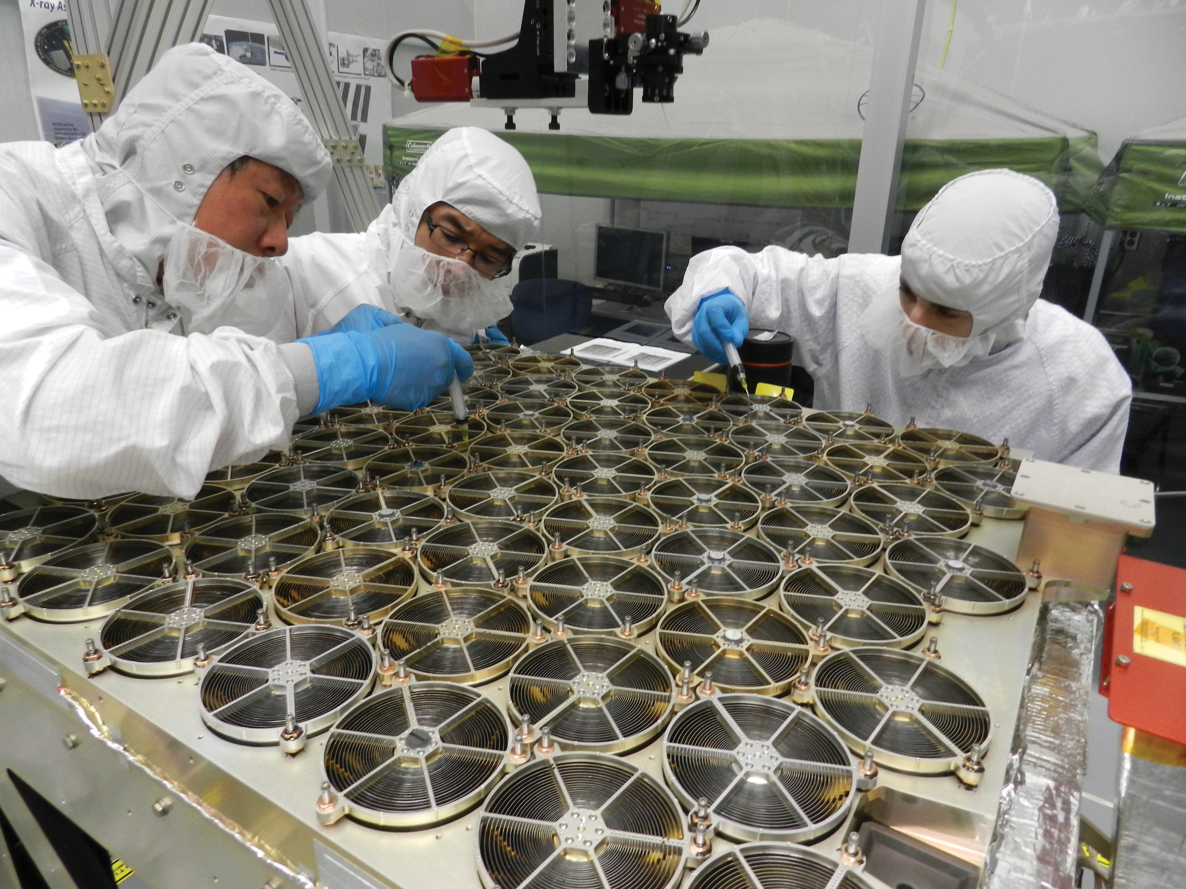


When the team behind NASA’s Neutron star Interior Composition Explorer (NICER) developed their astrophysics experiment for launch to the International Space Station, they faced unique challenges. Researchers needed to create new X-ray technologies to best study the glowing cinders left behind when massive stars explode as supernovae and form neutron stars. What the NICER scientists never expected was that the technologies they developed for overcoming these challenges in space could lead to new advances in medicine.
In 2017, NICER joined other astrophysics experiments such as the Japan Aerospace Exploration Agency’s (JAXA’s) CALET and MAXI and NASA’s AMS-02 in using the outside of the space station as a unique perch for studying the universe.
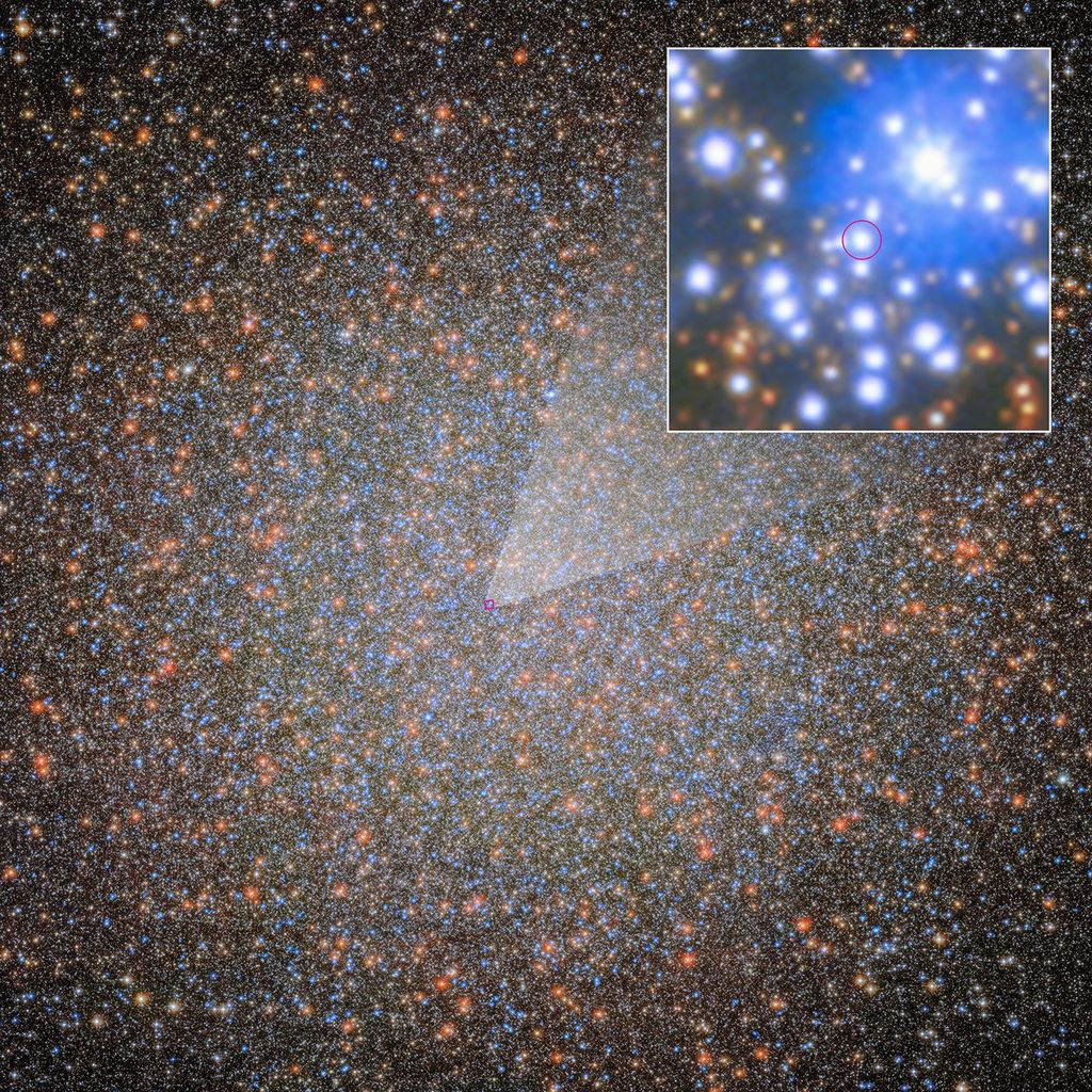
“We designed the mission to study neutron stars because they are really fantastic objects. They represent the densest matter that we know of in the universe. If you could squeeze a neutron star any further, it would implode into its event horizon and become a black hole,” said Keith Gendreau, NICER principal investigator at NASA’s Goddard Space Flight Center in Greenbelt, Maryland. “We do not know how matter behaves at these super-high densities because we cannot reproduce them in any experiment here on Earth or in the solar system.”
NICER’s observations of neutron stars have already led to numerous findings. In 2019, NICER detected a sudden spike of X-rays caused by a thermonuclear flash on the surface of a pulsar, a rapidly spinning neutron star. It also provided the first ever surface map of hot spots on a pulsar’s surface in the same year. In 2021, the telescope helped scientists discover that matter in the heart of a neutron star is less squeezable than some physicists predicted, NICER’s key science goal.
Scientists from around the globe are now using the payload to study everything from black holes to comets. For example, scientists used NICER to chart the environment surrounding J1820+070, a black hole about 10 times the Sun’s mass. That discovery made the cover of the January 2019 issue of the scientific journal Nature.
But before NICER did any of this, the research team worked on the ground to figure out how the experiment could provide accurate time-tag measurements of X-rays.
“NICER’s measurement capabilities that are unique involve timing. The instrument records to extremely high accuracy the time at which X-rays coming from the sky are received,” Gendreau said. “To verify that performance on the ground, we needed an X-ray generator that could produce X-rays at commanded times.”
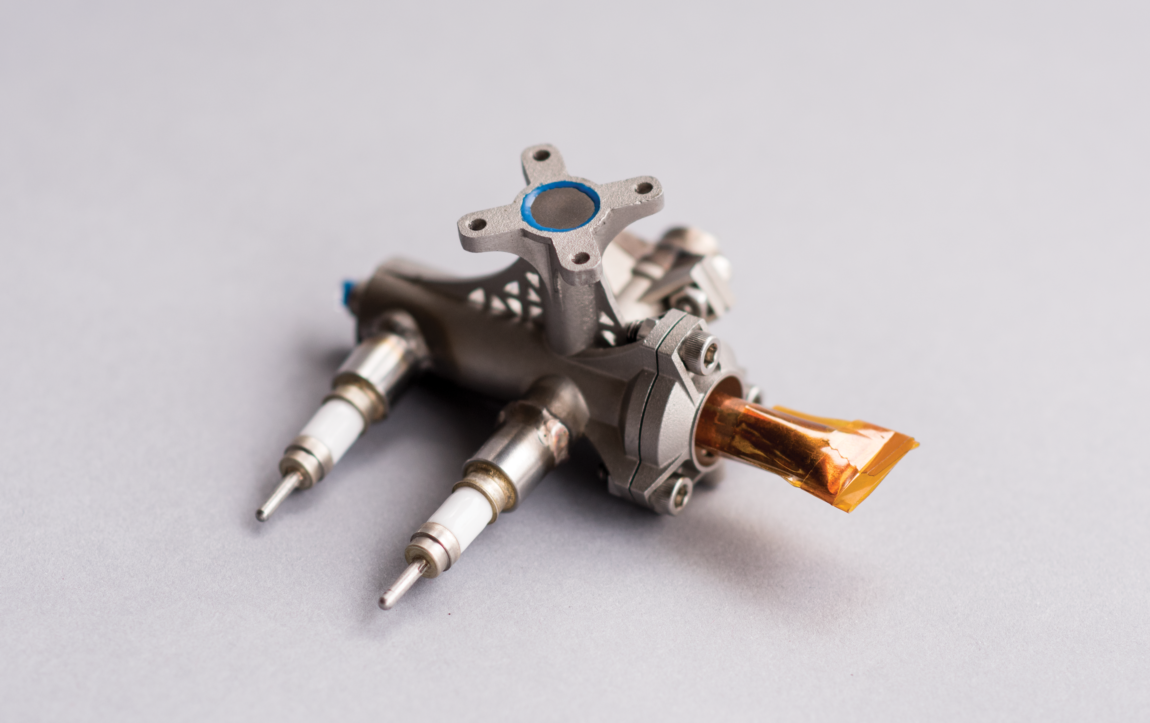
Traditional X-ray sources were limited in how quickly they could be turned on or off. The researchers needed to shorten that time from milliseconds to nanoseconds. To solve this challenge, the NICER team created and patented the Miniaturized High-Speed Modulated X-ray Source (MXS).
This also happened to be just what neuroradiologist Dr. Raj Gupta of Massachusetts General Hospital was looking for in his quest to improve computed tomography (CT) scans.
CT scans, formerly known as CAT scans, are obtained using rotating X-ray machines that image the body to provide important three-dimensional information to medical professionals. According to Harvard Health Publishing, more than 80 million CT scans are performed in the United States alone each year.
Traditional CT machines are large, heavy, and consume a lot of power, making them difficult to ship and hard to access in environments with few resources.
“Where I grew up, there is really no medical imaging. And even now there is very little penetration of advanced medical imaging there. Even simple X-rays are not available to about two-thirds of the world, according to the United Nations,” Gupta said.
Gupta lived in the small village of Malsisar in northwest India until he was nine years old. His desire to make medical technologies more accessible drove Gupta’s work to improve the devices. Coincidentally, what his research needed was similar to what the NICER team had created: a small, lightweight X-ray source that could turn on and off very quickly. When Gupta attended a talk put on by Gendreau, also the lead inventor of MXS, Gupta knew that it was something special. He saw the potential to increase the accessibility of CT scans, lower the radiation doses these procedures deliver to medical patients, and create new medical devices with potential use for space exploration. He reached out to Gendreau and the collaboration began.
“I never thought that when looking at some pulsars and neutron stars, I would find some technology that will be useful in my day-to-day work,” Gupta said.
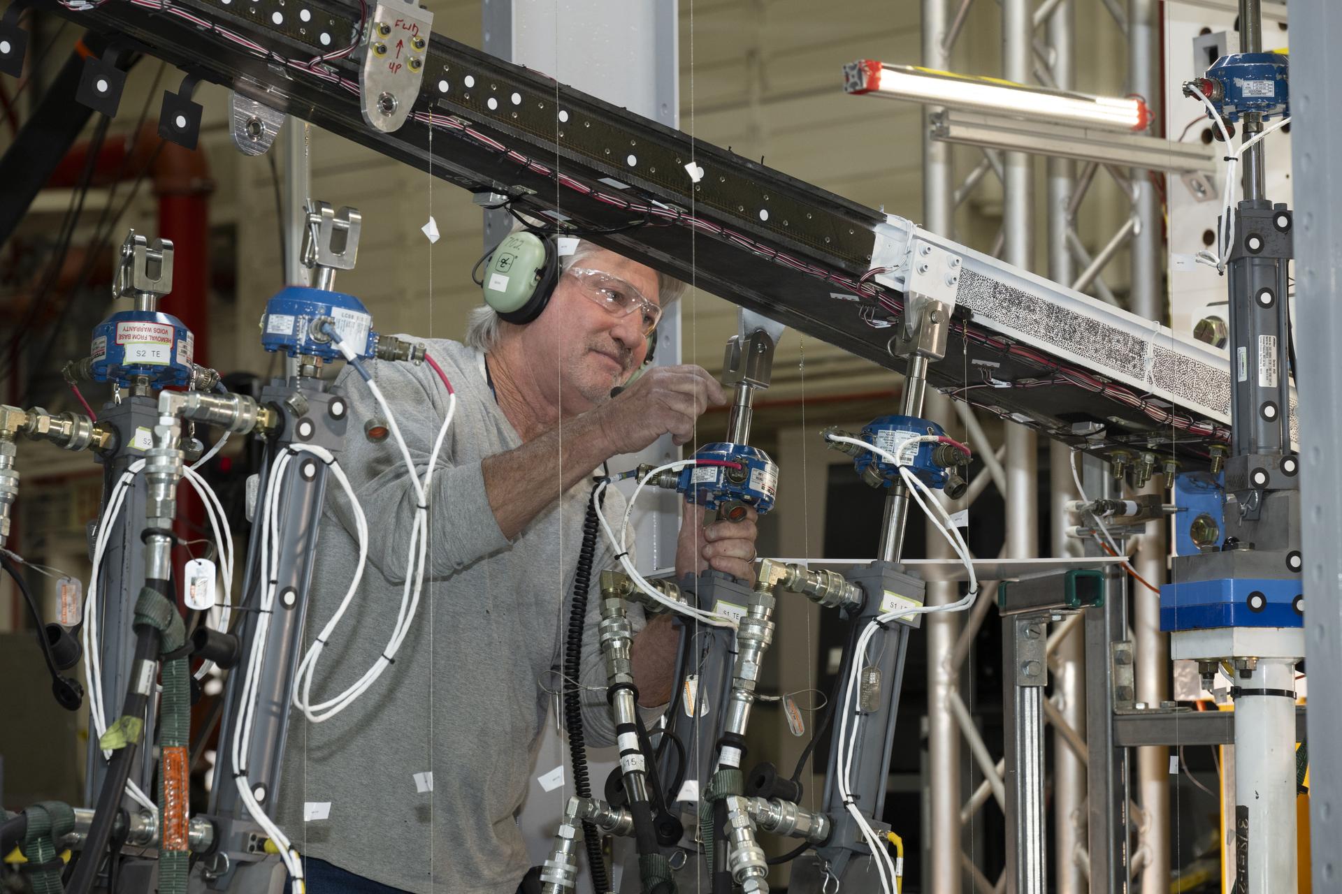
Rather than spinning the large X-ray machine around to capture a CT scan, Gupta worked with the NICER team to create a stationary ring of these new, small, modulated X-ray sources that can be mounted around the patient, firing when needed.
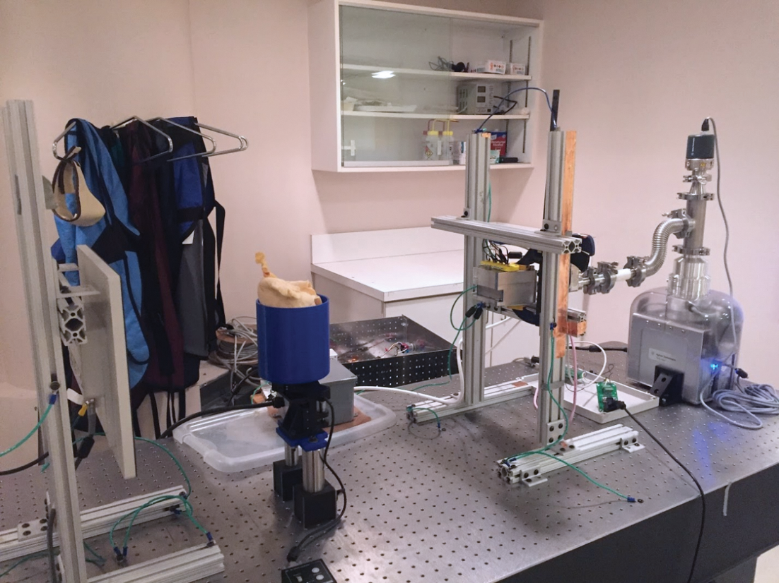
“If you have many X-ray sources around a person, then you can turn on the ones that are giving the doctor more information more often and those that are giving less information less often,” Gupta said.
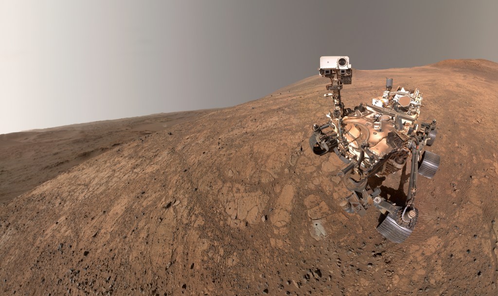
This technique can decrease the amount of radiation the patient is exposed to and enable better image quality for that level of radiation. Limiting radiation exposure is helpful for patients on Earth now and for potential future astronauts on their way to Mars.
“In space, because it is a high radiation environment to begin with, this is even more important,” Gupta said. “Currently, long missions take months, if not years, to complete. In that time, somebody could get sick or injured. There may be physicians aboard who can actually do something about it, but the traditional CT scanner is just not placeable in a space environment.”
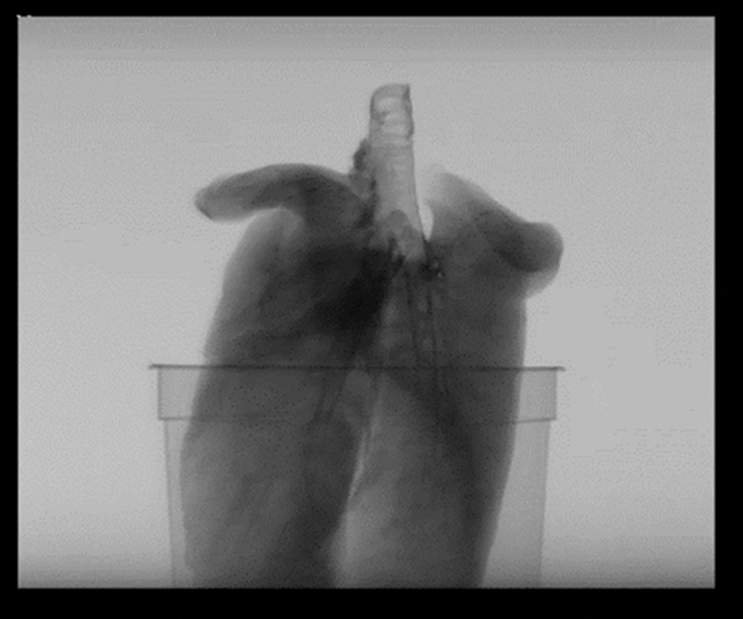
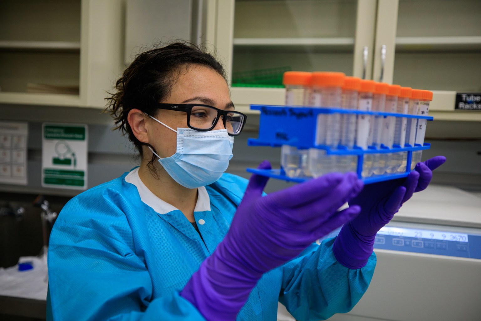
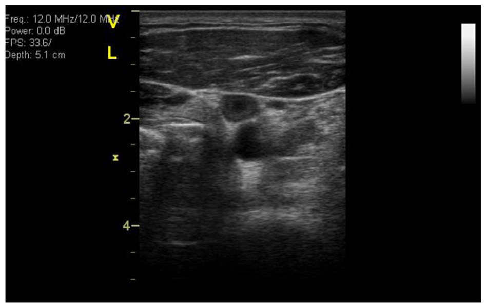
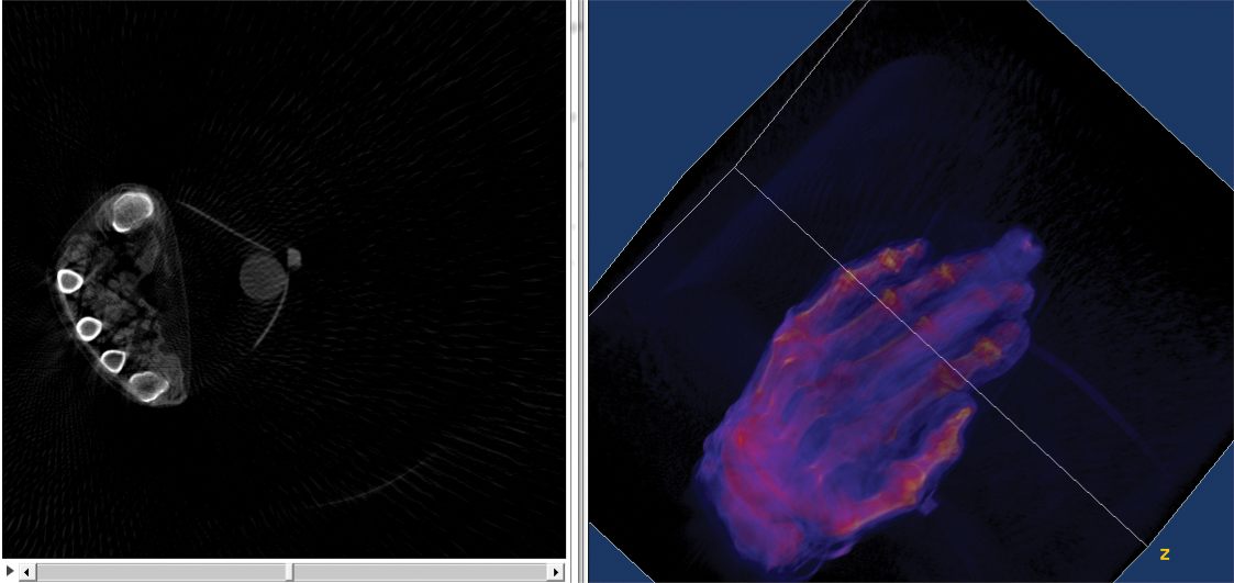
Gupta and his team have already performed successful test scans on pig lungs and human hands. They published their initial results in Nature Scientific Reports in 2018. Their next step is to increase the size and power of the setup for full-scale human testing.
“What has happened to the accessibility of consumer electronics has not happened to medical imaging, and ideas like this move the ball in that direction,” Gupta said.
He is hopeful that as more scientists from other fields study the work of NICER scientists, more medical benefits will arise.
“They have this method of taking X-rays and kind of concentrating them. They have essentially made a focusing system for X-rays that could be used in mammography,” Gupta said.
While this progress continues in the lab on Earth, NICER will continue staring up at the skies.
“There are lots of intriguing aspects about what goes on physically inside neutron stars, including superconductivity and superfluidity – all these weird quantum phenomena that are almost science fiction except that they’re not,” said Zaven Arzoumanian, NICER deputy principal investigator at Goddard.
Superconductivity and superfluids can be created in the lab, but only in tiny quantities. Neutron stars offer the rare opportunity to study quantities of superfluid that weigh as much as the Sun.
“We are exploring quantum phenomena that are happening on enormous scales, and understanding them can have technological relevance to us,” Arzoumanian said.
NICER’s continued work analyzing neutron stars and helping us better understand our universe may in turn lead to new ways of helping us back on Earth.